Corneal Nerve Analysis
Our Objectives
Cornea is the anterior clear part of the eye. It is the only location to evaluate the nerve in the body without any invasive technique.
We can acquire hundreds images of corneal nerve only by picturing the cornea of your eye with a camera called confocal microscopy.
There is the densest nerve plexus in the cornea called “sub-basal nerve plexus” that you can use this data to represent your systemic neuropathy such as Diabetes Mellitus, Parkinson’s Disease and more.
To our knowledge, there is no any validate automated program to analyze the corneal nerve at the same accuracy as manual counting. The current gold standard of corneal nerve analysis is manual segmentation by experts.
However, we are look forward to create an artificial intelligence for the best outcome of corneal nerve analysis. Anyone who are interested to collaborate with us can hand-in a resume to the contact details below.
Our mission is to provide medical personnels the best neuropathy data not only for your easier and more accurate practice, but also for the best quality of life for our patients.
Corneal Nerve Analysis
We provide the analysis of corneal nerve parameters shown below by using ImageJ program and NeuronJ plugin.
- Corneal Nerve Fiber Density (CNFD)
Length of all nerve fibers and branches per square millimeter.
Length of all nerve fibers.
Number of branches emanating from each main nerve per square millimeter.
The ratio of the length of the curve to the distance between its ends.
[Template for corneal nerve analysis report]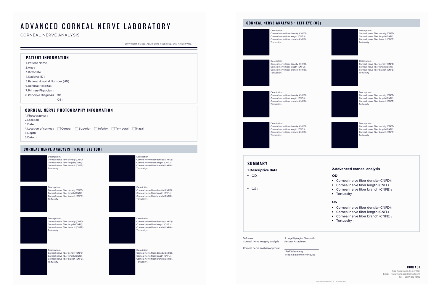
[Example of analysed IVCM]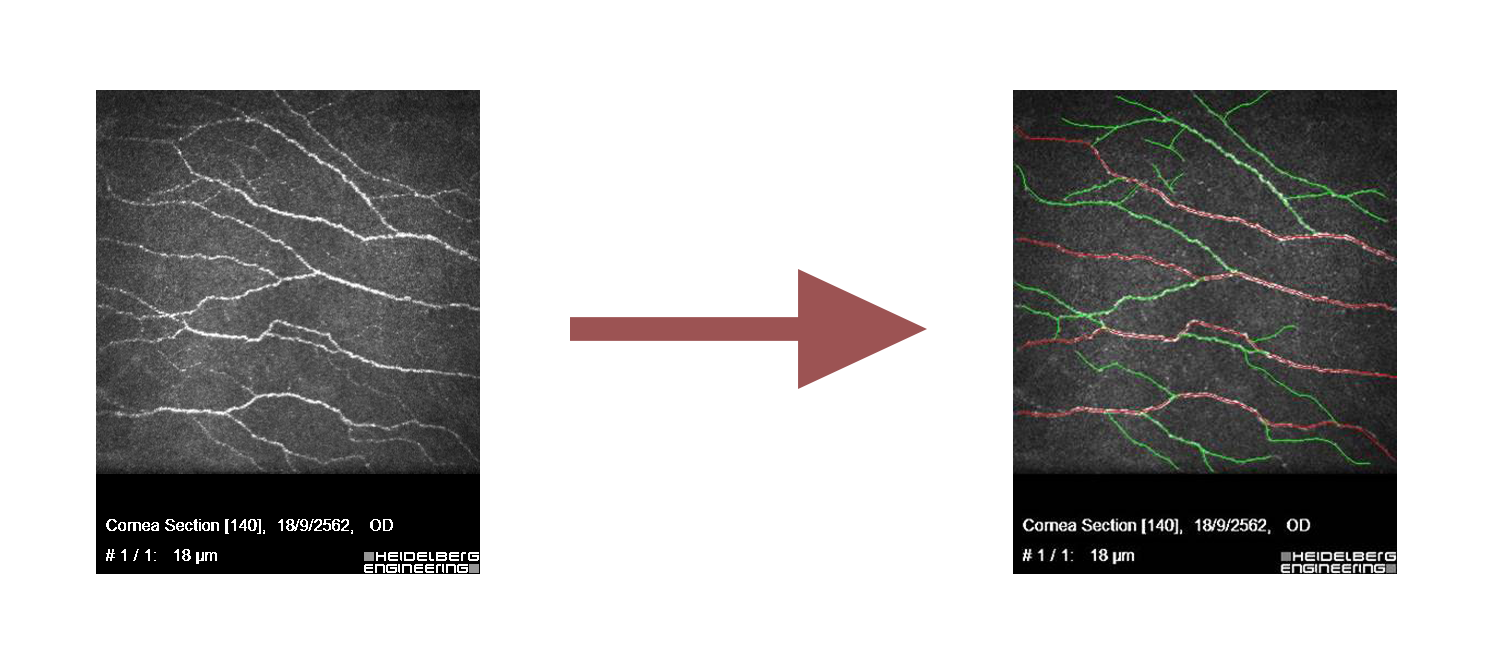
Contact

Sasi Yaisawang, M.D. FICO.
Corneal nerve analysis approver
Email: yaisawang.sasi@gmail.com